Clinical Details
- Two years and eight months old male presented with swelling in left zygoma region for two months
- Patient had history of fever, five months ago which lasts for three days
- Extra-oral examination revealed diffuse non-tender, non-compressible and non-fluctuant swelling of size approximately 2x1x1 cm in left zygoma
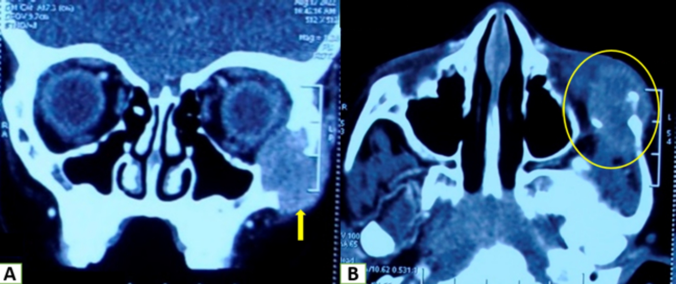
Radiographical Details
- Coronal and axial views are displaying a lytic expansile lesion involving the body of left zygoma and adjacent anterior zygomatic arch with cortical destruction.
Macroscopic Gross Details
- Two irregular brownish soft tissues (measuring 2.0x1.5x0.6 cm)
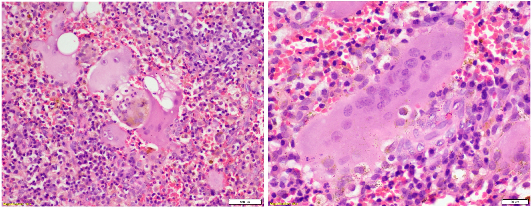
Microscopic Details
- Sheets of large multinucleated histiocytes (8 to 10 nuclei/cell)
- Giant cells showing emperipolesis and brown pigments within the cells
- Focal area of fibrosis
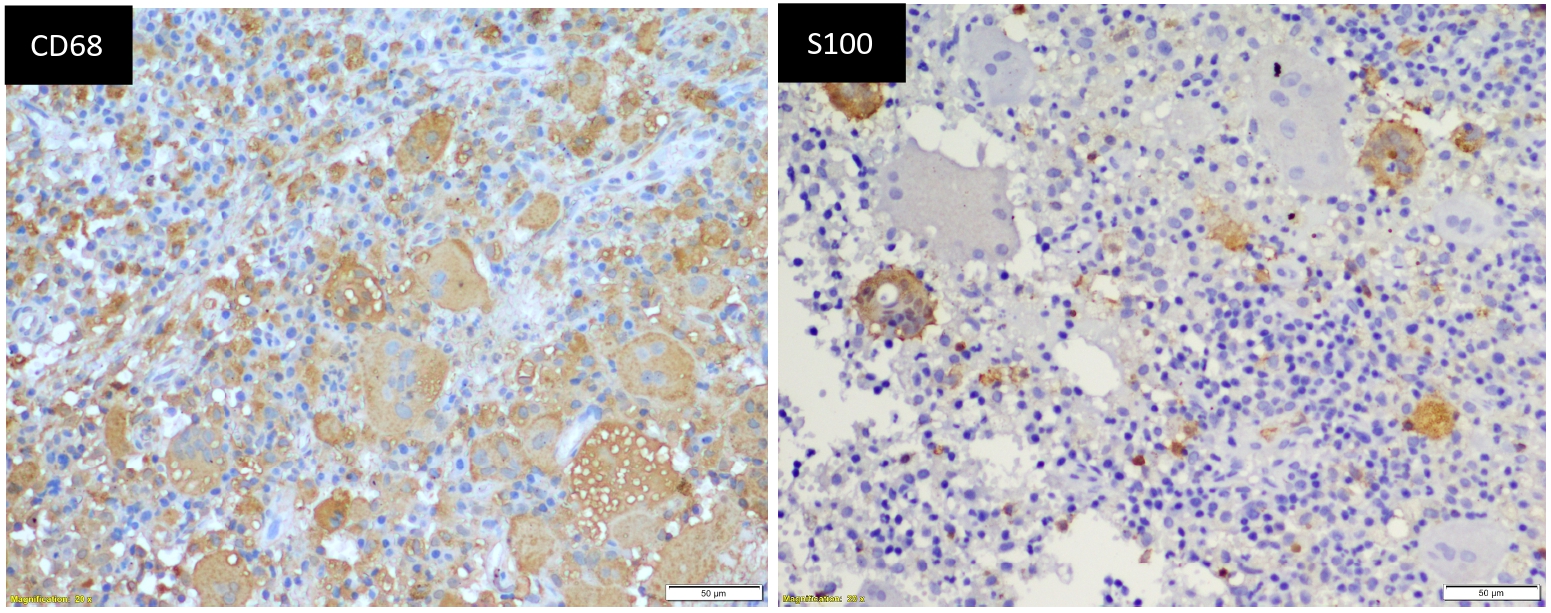
Immunohistochemical Details
- Multinucleated histiocytes showing immunopositivity for S100, CD68 and immunonegativity for CD1a.
Final Diagnosis
- Extranodal Rosai Dorfman disease of zygoma
Review of Literature
- Rare histiocytic disorder
- Usually presents as a painless lymph node adenopathy in adolescents and young adults
- Extranodal RDD most commonly involve the orbit, eyelid, upper respiratory tract, salivary gland, skin, bone, and testes. Bone involvement is rare (<10%)
- Approximately 22% cases of extranodal RDD has been reported in head and neck with nasal cavity and paranasal sinuses being the most frequently involved sites (~11%) in the pediatric population
- Microscopically, sheets of large histiocytes sometimes show multinucleation similar to our case. These cells show emperipolesis which is a non-destructive phagocytosis of lymphocytes and erythrocytes, which is the hallmark of the disease and is diagnostically helpful finding but not specific to RDD